


Case of the Month with Dr. Tommy Fung
Straumann BLX for Immediate Placement in Posterior Mandibular Area with Sealing Socket Abutment
Key Points:
Immediate implant placement is one of the hot topics in implant dentistry. The benefit of immediacy is to improve patient’s overall treatment perception and quality, in terms of the reduced number of visits and healing/handicapped period, without jeopardizing long term stability(Bhola, Neely, & Kolhatkar, 2008).
Careful examination of three dimensional imaging before extraction effectively guides clinicians to proper case selection and treatment planning(Smith & Tarnow, 2013).
An implant with tapered core and enhanced thread, can offer a secure grip into the socket wall after tooth extraction. Optimum primary stability is achieved in this challenging situation. The use of an implant with chemically modified surface further speeds up the process of new bone apposition without compromising the quality of osteointegration(Buser et al., 2004).
Initial Situation Description:
• A 41-year-old female requested a consultation. Tooth 36 was found with a distal caries with subgingival extension. The restorability was guarded.
• Treatment proposed including root canal treatment, crown lengthening and full contour crown placement for 36, was discussed with the patient.
• Primary examination including CBCT was taken to examine the local situation.
• Patient elected for extraction and replacement with implant after consideration of her time and need.
• Immediate implant placement was planned to allow better adaptation to the patient’s tight business schedule.
• Hygenic phase was completed with scaling and oral hygiene instruction.
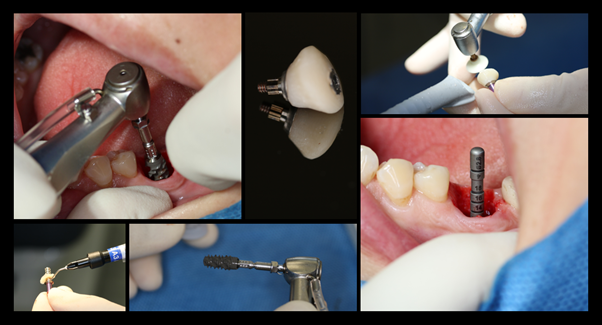
Surgical Procedure:
• Atraumatic extraction with sectioning and careful elevation of tooth roots were accompanished with the maintenance of intact socket wall.
• Implant (Straumann SLActive, BLX 5.5 x 12mm) was placed at correct 3D position at the interradicular septum.
• Initial primary stability of > 35Ncm was achieved.
• The space between the implant and socket was filled with particulate xenograft.
• A temporary abutment was modified with flowable composite, contoured according to the socket size and shape.
• The socket sealing abutment was further polished before tightening onto the implant ( >15Ncm ), covering the grafted area.
• Later, working impression with open tray technique was taken with a customized impression coping.
• Full milled zirconia crown with an exact copy of the emergence profile was connected and tightened to 35Ncm. Access screw hole was sealed with teflon tape and resin composite.


Discussion:
Immediate implant placement with conventional loading protocol is clinically documented in the posterior mandible and scientifically and clinically validated in the posterior maxilla(Zhou, Gallucci, Chen, Buser, & Hamilton, 2021). The survival rate was shown to be no different compare with delayed implant placement control(Ketabi, Deporter, & Atenafu, 2016). Within this context, the operator is confronted by two challenges. Firstly, the placement of an implant at the correct three dimensional position with adequate primary stability, regardless of the morphology of the sockets; and secondly, the proper transmucosal closure of the wound to cover and protect the grafted area.
It is vital to take into account the morphology of the socket in a three dimensional manner. Pre-operative assessment with cone beam CT gives a good estimate of the amount of bone left after extraction. The slope and curvature of the tooth roots, the dimension and radiodensity of the interradicular bone, and the presence or absence of pathological bone resorption around the area, provide hints whether the implant would be placed as planned, or drifted aside. Sometimes a partial removal of the interradicular bone provides a stable base for site preparation(Fugazzotto, 2002). Bur slipping could be avoided by pre-drilling at this ideal position with a needle bur or a sharp round bur. Xenograft was placed in the fixture-socket gap as a bone substitute material. It was shown to result in less horizontal buccal bone resorption than immediate placement of implant without bone grafting(Zaki, Yusuf, El-Khadem, Scholten, & Jenniskens, 2021).
Sealing socket abutment is a suitable tool for wound coverage after posterior immediate implant placement. Like the case of immediate restoration at the anterior area, it provides and maintains support to the soft tissue in the transmucosal zone. The intended low profile outline effectively avoids the potentially detrimental immediate-loading effect(Finelle, Popelut, Knafo, & Martin, 2021). This technique is also designed to provide a streamlined clinical workflow such that the anatomical morphology of a molar is maintained and the biocopy transferred throughout the surgical and restorative phase of the treatment(Finelle, Sanz-Martin, Knafo, Figue, & Popelut, 2019). The final emergence profile of the implant prosthesis resembles the original natural molar.
Conclusion:
Immediate implant is a double-edged sword. The actual anatomy of the freshly extracted socket, and the corresponding surgical technique utilized, dictate the overall treatment outcome. Straumann BLX implant with its self-cutting thread and fully tapered design, improves implant stability in this especially unfavorable condition. It certainly provides added value to the handling of cases of immediacy. After all, careful and accurate execution of the surgery cannot be overemphasized. It is crucial to weigh the immediate short-term benefit over the long-term clinical result.
Reference:
Bhola, M., Neely, A. L., & Kolhatkar, S. (2008). Immediate implant placement: clinical decisions, advantages, and disadvantages. J Prosthodont, 17(7), 576-581. doi:10.1111/j.1532-849X.2008.00359.x
Buser, D., Broggini, N., Wieland, M., Schenk, R. K., Denzer, A. J., Cochran, D. L., . . . Steinemann, S. G. (2004). Enhanced bone apposition to a chemically modified SLA titanium surface. J Dent Res, 83(7), 529-533. doi:10.1177/154405910408300704
Finelle, G., Popelut, A., Knafo, B., & Martin, I. S. (2021). Sealing Socket Abutments (SSAs) in Molar Immediate Implants with a Digitalized CAD/CAM Protocol: Soft Tissue Contour Changes and Radiographic Outcomes After 2 Years. Int J Periodontics Restorative Dent, 41(2), 235-244. doi:10.11607/prd.4579
Finelle, G., Sanz-Martin, I., Knafo, B., Figue, M., & Popelut, A. (2019). Digitalized CAD/CAM protocol for the fabrication of customized sealing socket healing abutments in immediate implants in molar sites. Int J Comput Dent, 22(2), 187-204.
Fugazzotto, P. A. (2002). Implant placement in maxillary first premolar fresh extraction sockets: description of technique and report of preliminary results. J Periodontol, 73(6), 669-674. doi:10.1902/jop.2002.73.6.669
Ketabi, M., Deporter, D., & Atenafu, E. G. (2016). A Systematic Review of Outcomes Following Immediate Molar Implant Placement Based on Recently Published Studies. Clin Implant Dent Relat Res, 18(6), 1084-1094. doi:10.1111/cid.12390
Smith, R. B., & Tarnow, D. P. (2013). Classification of molar extraction sites for immediate dental implant placement: technical note. Int J Oral Maxillofac Implants, 28(3), 911-916. doi:10.11607/jomi.2627
Zaki, J., Yusuf, N., El-Khadem, A., Scholten, R., & Jenniskens, K. (2021). Efficacy of bone-substitute materials use in immediate dental implant placement: A systematic review and meta-analysis. Clin Implant Dent Relat Res, 23(4), 506-519. doi:10.1111/cid.13014
Zhou, W., Gallucci, G. O., Chen, S., Buser, D., & Hamilton, A. (2021). Placement and Loading Protocols for Single Implants in Different Locations: A Systematic Review. Int J Oral Maxillofac Implants, 36(4), e72-e89. doi:10.11607/jomi.8750
