


Download Documents
Durable Cementation of Lithium- Disilicate Crowns and Veneers
24 Mar 2021
Lithium-disilicate has long been a reliable standard for indirect restorations, with its superb dimensional stability and precision finishing. In particular with the advancement of minimally invasive restorations and no-preparation veneers, Lithium-discilicate opens up great possibilities, but the cementation technique and material becomes even more critical for the final outcomes. The evolution of cements and cementation techniques can therefore be a key parametre in minimally invasive restorations. Further to the cement mechanical properties and influence in the aesthetics, the addition of bio-interactive or regenerative qualities can significantly increase the options for the clinician. Read about a calcium silicate–based dual-cure resin cement with calcium and fluoride release capabilities in the case report by Dr. Jack D. Griffin!
Abstract: Clinicians have many restorative options from which to choose. For indirect restorations, the selection of cementation technique is critical to a successful outcome. One emerging trend in dentistry today is the development of materials that have bio-interactive or regenerative qualities. This case presentation reports on the use of a calcium silicate–based dual-cure resin cement that has shown to be effective for use with full-coverage lithium-disilicate restorations due to not only its excellent clinical characteristics but also calcium and fluoride release capabilities. The case also includes conservative lithium-disilicate veneer restorations.
Many viable material choices are available in restorative dentistry today. Despite the ever-growing arsenal of newer materials and techniques, clinicians are often reluctant to change from using those materials and methods with which they are most familiar and have had success. Above all else, clinicians want predictable, repeatable results. Restorative dental materials have advanced to meet the increasingly bio-friendly and metal-free esthetic demands of the public today. Contemporary materials are expected to have, not a negative, but a positive effect on living tissues. An aversion to metal, the avoidance of potential allergies, and the systemic effect of dental materials all drive the dental profession to be more biologically tolerant.
Versatile Predictability
When working with indirect restorative materials, clinicians require consistency in esthetics, functionality, durability, and patient comfort. Zirconia and lithium disilicate have become dominant materials in modern metal-free dentistry. They are commonly used for restorations ranging from conservatively prepared veneers to full-coverage crowns to full-arch prostheses. Factors such as occlusion, parafunctional habits, esthetics, and biological effects influence a clinician’s choice of indirect materials. The success of lithium disilicate, specifically IPS e.max® (Ivoclar Vivadent, ivoclarvivadent.com), has been well documented, and it has become one of the most versatile esthetic materials in dentistry. The dental profession has had more than 15 years of clinical performance and numerous studies to fairly evaluate this material, its clinical properties, and durability. Success has been observed in both full-coverage and conservative restorations as well as in anterior and posterior situations. High translucency imparts vitality to a restoration and is important to its esthetic success.9 The esthetic predictability of lithium disilicate, particularly when used in conservative anterior preparations, makes it a popular cosmetic material choice. Lithium disilicate is available in several different levels of opacities. The opacity should be chosen on a case-by-case basis depending on preparation shade, thickness of the restoration, and desired final shade.
Modern Cementation
Cementation technique is critical to restoration success, and clinicians have many materials and methods from which to choose: cement or bond, light cure or chemical cure, acid-etch or no etch, bonding agent or self-adhesive, silane or sandblasting, etc. Retention is but one of many factors influencing cement choice. Other factors include long-term physical properties, color predictability, ease of clean-up, and the luting material’s compatibility with the restoration material. Self-adhesive resin cements work well with retentive preparations, but light-cure resin cements with separate steps for phosphoric acid-etching and adhesion provide better long-term results with preparations that have less-than-ideal retention.
Light-cure-only resin cements are often used with conservative anterior restorations because of their longer working time, predictable clean-up, excellent esthetics and color stability, and high strength. Transparent light-cure resins have been demonstrated to yield superior color stability when compared to luting materials with more color or that are dual cure. Full-coverage restorations allow for greater diversity in cementation choices. Self-adhesive dual-cure resin cements are popular with full-coverage restorations because of their simplicity and predictable results. They provide a moderate bond to dentin and good esthetics. A newer emphasis has been placed on cements that are bio-interactive or regenerative in nature. Bioavailable ions generate an alkaline pH environment, neutralize acid, and ultimately promote healing, which may result in a less sensitive, longer-lasting restoration. The cement responds to the oral environment to provide therapeutic ions. Calcium silicate–based dual-cure resin cements have shown to be an effective choice for full-coverage lithium disilicate because of excellent clinical characteristics along with calcium and fluoride release. The following case report demonstrates the use of an ionreleasing self-etch, self-adhesive calcium-silicate resin cement with layered lithium-disilicate crowns and a translucent lightcure resin cement for placement of lithiumdisilicate veneers.
Crown and Veneer Case
A 61-year-old patient presented with her tooth No. 9 crown loose, and she could actually remove it (Figure 1). Many years earlier the patient had had endodontic surgery, an apicoectomy and retrograde filling to repair a failing silverpoint obturation, and was now symptom free. The silver point remained in the tooth and showed no lesions on radiograph. Tooth No. 8 had an unbonded porcelain veneer that had come off “several times” before. She wanted to replace the restorations on the central incisors with ones that would be a “lighter color” and to bleach her other teeth (Figure 2). A full series of photographs was taken and reviewed with the patient. Occasionally, in the author’s experience, patients might want to choose a “lighter color” for their restorations and attempt to bleach their other teeth until they match. After some discussion and patient education, the patient accepted a plan calling for lithium-disilicate porcelain restorations on teeth Nos. 5 through 12, which included full coverage on the central incisors and conservative veneer preparations on the other teeth. A full-coverage restoration for tooth No. 8 was chosen because the existing preparation was primarily in dentin, there was a history of veneer failure, and doing so would provide the laboratory with symmetrical consistency in restoration thickness. Another full series of photographs was taken, a mock-up was done, and a temporary impression matrix was made. The patient chose a final VITA 3D shade of .5M1 (VITA, vitanorthamerica.com). Preparations were done using a course diamond (NeoDiamond®, Microcopy, microcopydental.com), with 1.5 mm occlusal clearance and chamfer margins. On teeth Nos. 5 through 7 and 10 through 12, 0.5 mm veneer preparations were executed with a finishing diamond, staying in enamel in all places except abfraction areas at the gingival margins. Preparation corners were rounded to reduce internal stresses on the restorations.


Fig 1. Patient with failed veneer No. 8 and dislodged crown No. 9. Fig 2. Treatment goals were to replace restorations on the central incisors and lighten color throughout the dentition. Fig 3. After restoration removal and veneer preparation, shade photographs were taken. Fig 4. Silver point was removed from coronal portion of tooth No. 9, and build-up was done with dual-cure material resulting in less darkness on the tooth.
Shade photographs were taken on moist teeth with shade tabs on the same plane as the teeth (Figure 3). Because of the dark gray color of the endodontically treated tooth No. 9, the silver point was removed from the coronal portion of the tooth and replaced with a dual-cure build-up material (CoreFlo™ DC, BISCO, bisco.com), shade Natural/A1 (Figure 4). Communicating the final preparation shades accurately to the laboratory was critical in enabling the lab to choose the most translucent shade of lithium disilicate that would mask the preparation and achieve the final color desired. Conventional impressions were taken with a polyvinyl siloxane material (Panasil® , Kettenbach, kettenbach-dental. us) in a stock tray and sent to the laboratory with all photographs, the bite registration, and a dentofacial analyzer index (Kois Dentofacial Analyzer, Panodent, panodent. com). Temporary restorations were made for the central incisors, 2% chlorhexidine (Cavity Cleanser™, BISCO) was painted on the teeth, and the restorations were placed with a temporary cement (ZONEfree™, Pentron, pentron.com). The veneer preparations were temporized via spot-etching and lock-on technique without the use of a bonding agent. All records were sent to the laboratory and all case photographs were uploaded to the lab portal. After 5 days, the patient returned for evaluation of color, length, and function with the temporaries. Slight adjustments were made, polishing was done, photographs were taken, and desired changes were
communicated with the lab.
Cementation for Crowns
The layered lithium-disilicate restorations were returned from the laboratory having been hydrofluoric acid-etched and silanated, and ready for cementation. The temporaries were removed, the teeth were cleaned with flour pumice, and the restorations were inspected in place. No adjustments were needed in this case. All restorations were rinsed, cleaned with a restoration cleaner (ZirClean®, BISCO) to remove contaminants. The two crowns were cemented with a self-etch, self-adhesive calcium-silicate resin cement (TheraCem® , BISCO) and held in place for about 1 minute; excess cement was easily removed (Figure 5). After complete clean-up light-curing was done at the margins. In the author’s experience, the calcium and fluoride release, easy clean-up, and physical properties make this cement an excellent choice for routine lithium-disilicate full-coverage placement. This self-etch, self-adhesive cement is available in a “natural” shade and provides firm bond to dentin and most modern dental materials such as etched lithium disilicate and zirconia. The high calcium content in this calcium and fluoride ion-releasing cement gives the material a slightly opaque, white appearance but does not seem to influence the final restoration shade in routine indirect restorations.
Cementation for Veneers
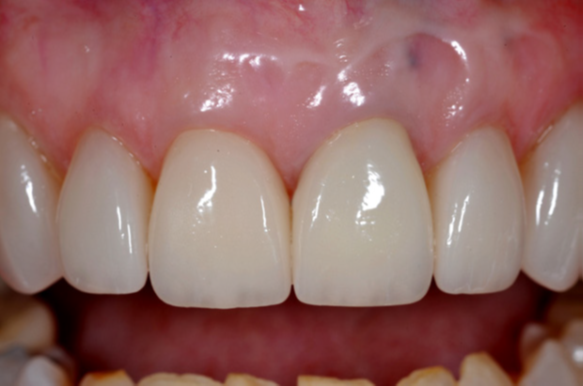
The intaglio surfaces of the veneers after re-silanation were thoroughly dried, and a universal dentin bonding agent (All-Bond Universal® , BISCO) was applied and airthinned. The teeth were isolated with retractors, etched with 37% phosphoric acid (Etch-37™ with BAC, BISCO) for 15 seconds, and rinsed (Figure 6). The universal adhesive was applied in several applications, allowed to sit for 20 seconds, air-dried with an air syringe for 10 seconds, and then lightcured for 10 seconds (Figure 7). After material was extruded from the tip onto a napkin, the translucent light-cure resin cement (Choice™ 2, BISCO) was applied to each tooth, and the veneers were placed ensuring that excess material extruded from all margins (Figure 8). The excess material was brushed away and all restorations were flossed while being held securely in place. Along with longterm color stability, the light-cure-only material allowed for thorough clean-up and an efficient cementation process. With each veneer being held in place, the restorations were cured with an LED light. Minor adjustments were made with a finish diamond, and the restorations were polished. The patient was scheduled for a follow-up appointment for adjustments as needed, cement clean-up, final photographs, and to place direct composite on the facial surfaces of the second bicuspids for enhanced blending. The softtissue response at 6 months was excellent for both the fullcoverage restorations and the veneers (Figure 9). The blending of materials, despite varied restoration thicknesses, was acceptable, and the patient enthusiastically approved of the final color shade (Figure 10).


Fig 7. Several coats of universal bonding agent were applied, air-dried, and light-cured. Fig 8. Light-cure-only cement was applied to the teeth, veneers were placed, clean-up was completed, and light-curing was done. Fig 9. Soft-tissue response was excellent, as was long-term prognosis. Fig 10. Good opacity of the selected lithium disilicate resulted in a consistent final shade.
Conclusion
As materials for indirect restorations continue to advance, biologically friendly cements can help generate a less sensitive, longer-lasting restoration through calcium and fluoride release. As demonstrated in this case report, a calcium silicate–based dual-cure resin cement, offering excellent clinical characteristics, including easy clean-up, was an effective choice for full-coverage lithium-disilicate anterior restorations.
About the Author
Jack D. Griffin, Jr., DMD
Accredited Member, American Academy of Cosmetic Dentistry;
Diplomate, American Board of Aesthetic Dentistry;
Master, Academy of General Dentistry;
Private Practice, Lake St. Louis, Missouri